KOGO 2026
Spatial transcriptomics reveal distinct tumor microenvironment associated with clinical response in thymic epithelial tumors after neoadjuvant therapy
Background: Thymic epithelial tumors (TETs) are rare malignancies, mostly treated with surgical resection as the standard approach. However, recurrence rates remain high and immunotherapy can lead to immune-related adverse events, highlighting the need for improved therapies and predictive biomarkers. In this study, TET patients were administered three cycles of neoadjuvant immunochemotherapy prior to surgery. We aimed to characterize the tumor microenvironment (TME) to identify the association with treatment response.
Methods: We performed 18 single-cell RNA sequencing and 15 spatial transcriptomics on surgical specimens from 19 patients treated with neoadjuvant immunochemotherapy. Major pathological response (MPR) was defined as less than 10% viable tumor cells in resected specimens. Patients were stratified into three pathological response groups ⎯ thymic carcinoma (TC) with MPR, TC non-MPR, or thymoma (TM) non-MPR.
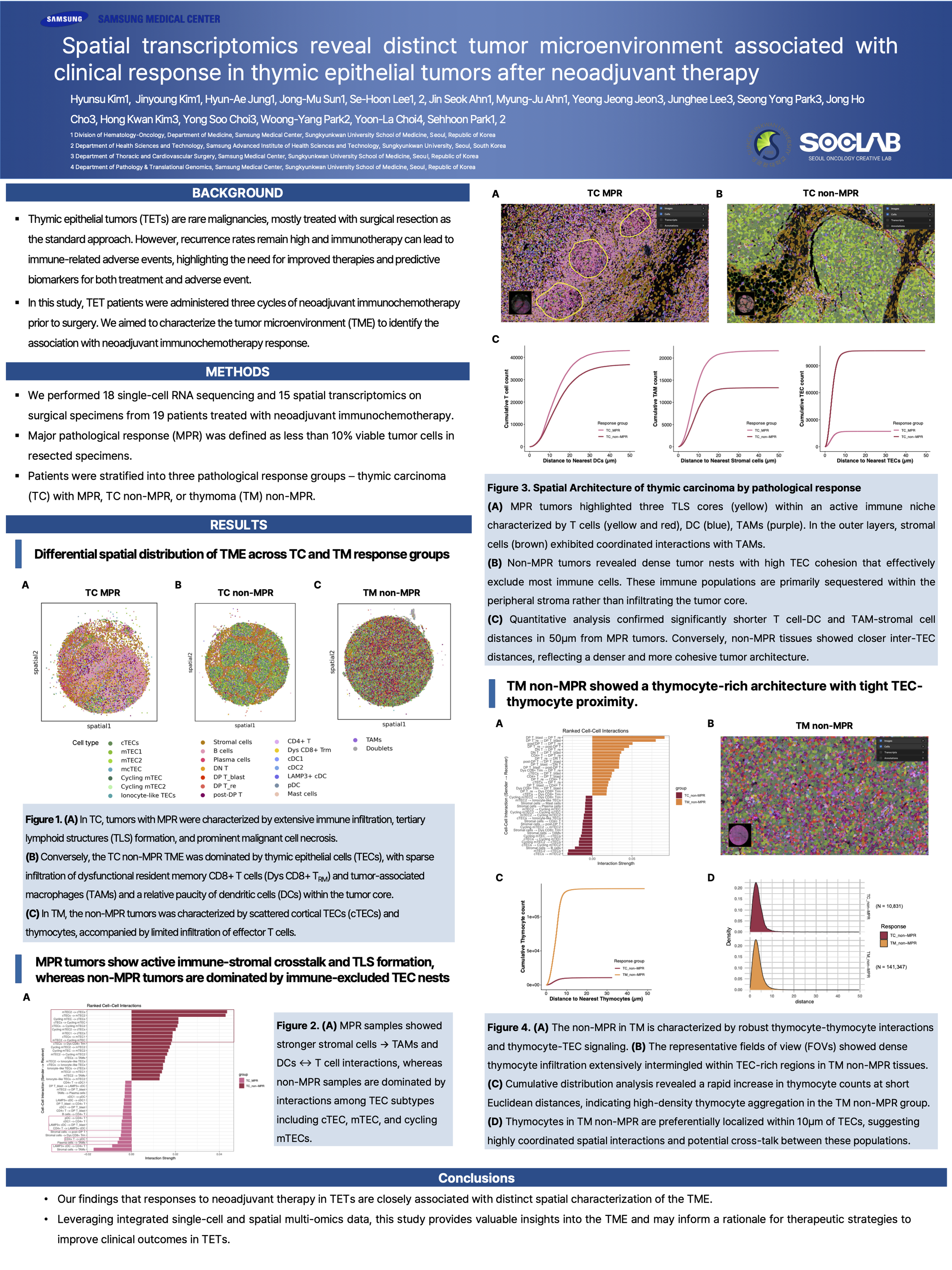
Results: Analysis of cell composition and ligand-receptor interaction networks revealed distinct TME differences according to pathological response group. In TC, tumors with MPR were characterized by extensive immune infiltration, tertiary lymphoid structures (TLS) formation, and prominent malignant cell necrosis. This immunologically hot TME featured robust interactions between lymphocytes and dendritic cells (DCs). Conversely, the TC non-MPR TME was dominated by thymic epithelial cells (TECs). Within the tumor core, these TECs orchestrated a localized immunosuppression by promoting the exhaustion of infiltrating CD8+ T cells and driving the polarization of tumor-associated macrophages into an M2-like state. This suppressive niche was further compounded by a scarcity of DC. In the context of TM, the non-MPR tumors was defined by scattered cortical TECs (cTECs) and thymocytes, alongside a sparse infiltration of effector T cells. Notably, cTECs maintained an immature TME by promoting thymocyte survival and retention while concurrently impairing the tumor-killing functionality of CD8+ T cells. Detailed cell-to-cell interaction results that further elucidate these TME dynamics will be presented.
Conclusions: Our findings that responses to neoadjuvant therapy in TETs are closely associated with distinct spatial characterization of the TME. Leveraging integrated single-cell and spatial multi-omics data, this study provides valuable insights into the TME and may inform a rationale for therapeutic strategies to improve clinical outcomes in TETs.