KOGO 2026
Longitudinal Tumor Microenvironment Analysis to Predict Immunochemotherapy Response in Salivary Duct Carcinoma
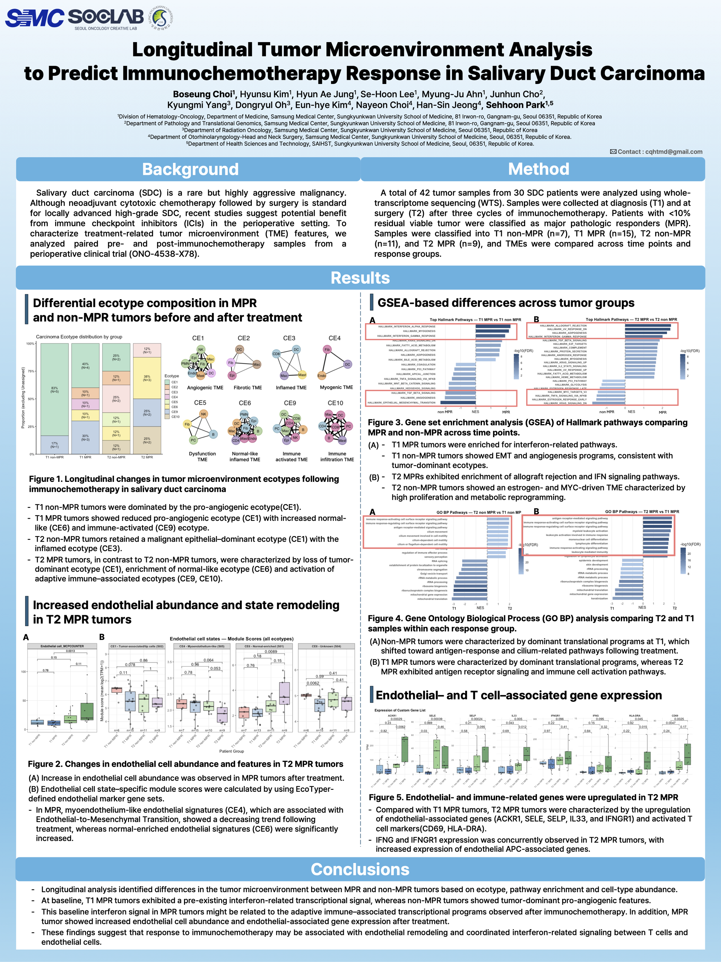
Background : Salivary duct carcinoma (SDC) is a rare but highly aggressive malignancy. Although neoadjuvant cytotoxic chemotherapy followed by surgery is standard for locally advanced high-grade SDC, recent studies suggest potential benefit from immune checkpoint inhibitors (ICIs) in the perioperative setting. To characterize treatment-related tumor microenvironment (TME) features, we analyzed paired pre- and post-immunochemotherapy samples from a perioperative clinical trial (ONO-4538-X78).
Method : A total of 42 tumor samples from 30 SDC patients were analyzed using whole-transcriptome sequencing (WTS). Samples were collected at diagnosis (T1) and at surgery (T2) after three cycles of immunochemotherapy. Patients with <10% residual viable tumor were classified as major pathologic responders (MPR). Samples were classified into T1 non-MPR (n=7), T1 MPR (n=15), T2 non-MPR (n=11), and T2 MPR (n=9), and TMEs were compared across time points and response groups.
Results : Cell-state inference revealed distinct TME profiles. At baseline (T1), non-MPR tumors exhibited a pro-angiogenic TME driven by malignant epithelial cells, consistent with EMT and angiogenesis pathways. In contrast, T1 MPR tumors showed a more immune-infiltrated TME with interferon-related signaling.
From the surgical samples (T2), direct comparison of MPR and non-MPR revealed distinct antitumor immune patterns, indicating robust T- and B-cell–mediated adaptive immunity in MPR, whereas non-MPR displayed chronic inflammatory signatures rather than effective antitumor responses.
Longitudinal comparison further highlighted divergent trajectories. In non-MPR tumors, T2 non-MPR tumors were inferred to retain malignant epithelial features with minimal immune cells; DEG analysis supported this with inflammatory responses at T2 and EMT programs at T1. Conversely, T2 MPR tumors were inferred to lose malignant epithelial signatures and show immune activation, consistent with T cell–mediated adaptive responses pathways, whereas T1 MPR displayed epithelial cell-cycle pathways.
Conclusion : In this study, we found that MPR tumors exhibited a pre-existing interferon-activated tumor microenvironment at baseline, which transitioned into an adaptive immune response after immunochemotherapy. These findings suggest that an interferon-primed TME is a prerequisite for effective post-treatment immune activation and may serve as a predictive biomarker for immunochemotherapy responsiveness.